
articles
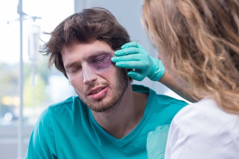
Urgent eye care encompasses prompt evaluation and treatment of sudden or severe eye-related issues, including foreign object removal, chemical exposure, corneal abrasions, sudden vision loss, eye trauma, acute glaucoma, chemical burns, and eye infections. Seeking immediate professional attention from an optometrist is vital to prevent further damage and preserve vision.
Common Eye Emergencies or Urgent Eye Care Appointments
Eye emergencies can manifest in various forms, and it is essential to be able to identify them quickly. Some common eye emergencies include:
Foreign Object in the Eye: Particles, debris, or small objects can become lodged in the eye, causing pain, redness, tearing, and potential damage to the eye's surface.
Corneal Abrasions or Scratches: Injuries to the cornea, such as abrasions or scratches, can cause severe eye pain, light sensitivity, and a feeling of something in the eye.
Sudden Loss of Vision: Any sudden and unexplained loss of vision requires immediate attention to determine the underlying cause and initiate appropriate treatment.
Eye Trauma or Blunt Force Injury: Injuries to the eye from impact, trauma, or accidents can lead to serious complications, including retinal detachment, hemorrhage, or intraocular foreign bodies.
Chemical Burns: Exposure to caustic substances or chemicals can cause serious damage to the eyes, resulting in pain, redness, and potential vision loss.
Eye Infections: Infections such as conjunctivitis (pink eye) can cause redness, discharge, and discomfort in the eyes.
Recognizing these symptoms and seeking urgent care can prevent further complications.
The Importance of Basic Red Eye Exams in Urgent Care
Red eye exams are a fundamental part of urgent eye care. They help identify the cause of redness and determine the appropriate treatment. Basic red eye exams involve a comprehensive evaluation of the eye, including examining the eyelids, conjunctiva, cornea, and iris. These exams aid in the diagnosis and treatment of various conditions, such as conjunctivitis, uveitis, dry eyes, and corneal abrasions.

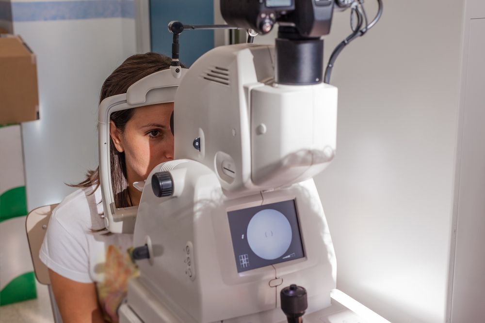
Optomap is an innovative new technology that gives eye doctors the ability to perform ultra-wide retinal imaging that is far superior to what can currently be achieved using conventional retinal imaging options. In contrast to conventional retinal imaging, Optomap captures at least 50% more of the retina in a single capture, and with Optomap’s multi-capture function, up to 97% of the retina can be viewed. This gives eye care professionals greater opportunity to monitor the health and condition of patient vision.
Why is Optomap important?
Optomap is another great preventative eyecare technology tool. By allowing your eye doctor to have a comprehensive view of your retina, they will be able to detect any developing eye diseases early on, before they have a detrimental impact on your vision and day to day life. Not only can Optomap detect eye conditions such as retinal holes, retinal detachment, macular degeneration and diabetic retinopathy, but it can also be used to identify some general health conditions such as cardiovascular disease, stroke and cancer.
What to expect from Optomap scanning
Optomap is a fast, painless and non-invasive procedure that is suitable for patients of all ages, even children and pregnant women. Many patients require their eyes to be dilated ahead of the scan and will be given eyedrops which will widen their pupils and make it easier for the camera to see the structures inside the eye. Pupil dilation is painless, but patients may feel more sensitive to light both during their Optomap scan and afterwards for up to 24 hours. You may also have slightly blurred vision for a few hours. Once your eyes are dilated, you’ll be sat down and asked to look into a small device that will take the pictures of your retina. A short flash of light will let you know that the image has been taken, and the entire imaging is over in just a few seconds. The results will be sent digitally to your eye doctor who will then evaluate them. The results will also be stored on your personal optical record for future information.
If you would like more information about what is involved in Optomap, or to schedule an appointment for this effective screening technology, please contact our eyecare team.

If you find it difficult to tell colors apart, you may be color blind. Color blindness, or color deficiency, is estimated to affect around 8% of men and about 1% of women, but for those affected, it can significantly impact the quality of their day-to-day life. Contrary to popular belief, being color blind doesn’t mean that you can’t see any color at all. Instead, patients simply struggle to differentiate between certain colors. The vast majority of people who are color blind find it impossible to tell the difference between varying shades of red and green. You may hear this referred to as red-green color deficiency. However, this doesn’t only mean that they mix up red and green. They can also mix up colors that have some green or red light as part of their whole colors, for example purple and blue. This is because they are unable to see the red light that forms part of the color purple.
As you can probably imagine, this type of visual impairment can be a problem for things like traffic lights, taking medications and even looking at signs and directions. For example, someone who is color blind may find that the green on a traffic light may appear white or even blue.
EnChroma lens technology is specifically designed to counteract red-green color deficiency and enable patients to better identify the difference in these colors or shades. They do this by selectively filtering out the red and green wavelengths of light at the exact point where the color sensitivities overlap before hitting the retina, creating far greater contrast between the colors so that the patient can distinguish between them successfully. Most cases of color blindness respond well to EnChroma’s innovative spectral lens technology, giving patients the ability to experience life in bright, vibrant technicolor.
EnChroma lenses are made from leading edge, Trivex material, and this helps to give them the best possible quality and clarity of vision. These lenses are also extremely light, strong and offer patients 100% protection against UV light, helping to keep your eyes healthy as well as improving your vision.
If you or someone you know is color blind or color deficient and could benefit from EnChroma lenses, contact us today to learn more about how they can help!
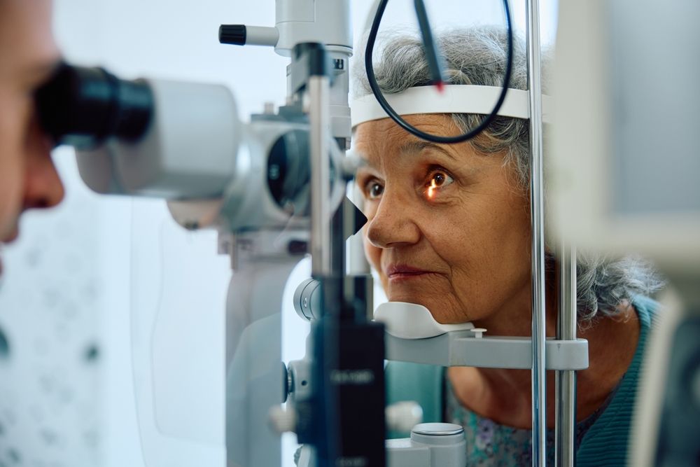
When it comes to maintaining your eye health, regular check-ups and screenings play a crucial role. One such screening method that has revolutionized the field of optometry is retinal imaging testing. This non-invasive procedure allows optometrists to capture detailed images of the retina, providing valuable insights into the overall health of your eyes.
The Importance of Retinal Imaging Testing
By examining the retina, which is the thin layer of tissue at the back of your eye responsible for capturing light and transmitting visual signals to the brain, optometrists can gain valuable insights into your eye health. Retinal imaging testing allows for the early detection of various eye conditions even before noticeable symptoms occur. This early detection is crucial as it enables prompt treatment and intervention, potentially preventing irreversible vision loss.
Advanced Retinal Imaging Technology
Retinal imaging testing has been made possible by the advancements in technology, specifically Optical Coherence Tomography (OCT) and Optos imaging. OCT is a non-invasive imaging technique that uses light waves to capture high-resolution cross-sectional images of the retina. It provides detailed information about the layers of the retina, helping eye doctors identify and monitor various eye conditions.
Optos imaging utilizes ultra-widefield retinal technology to capture a panoramic image of the retina. This technology allows for a more comprehensive view of the retina, including the periphery. Dilating drops are not necessary with Optos, making the process more convenient for patients.

Dry eye is a common condition that occurs when your eyes do not produce enough tears or when the tears evaporate too quickly. This can result in discomfort, irritation, and a gritty sensation in the eyes. Understanding the causes and symptoms of dry eye is crucial for finding effective treatment options. Tyrvaya offers a breakthrough solution for dry eye relief.
What Causes Dry Eye?
There are several factors that can contribute to the development of dry eye. The meibomian glands are responsible for producing the oily component of the tear film, which helps prevent evaporation of tears and maintains a smooth ocular surface. Meibomian gland dysfunction occurs when these glands become blocked, leading to a decrease in the quantity and quality of the meibum. This can result in evaporative dry eye, discomfort, and inflammation of the eyelid margins.
Another common causes is age. As we get older, our tear production tends to decrease, making us more prone to dryness. Hormonal changes, such as those that occur during menopause, can also affect tear production and lead to dry eye.
Environmental factors can play a role as well. Dry or windy climates, air conditioning, and excessive screen time can all contribute to dry eye. Additionally, certain medications, such as antihistamines and antidepressants, can cause dryness as a side effect.
Other underlying health conditions, such as autoimmune diseases like Sjogren's syndrome or rheumatoid arthritis, can also contribute to dry eye. In these cases, the body's immune system mistakenly attacks the tear glands, leading to reduced tear production.
Medical eye exams are comprehensive evaluations of the health and function of the eyes, essential for maintaining optimal vision and preventing potential eye conditions. Regular medical eye exams are fundamental for proactive eye health management and ensuring ongoing visual wellness.
What is a Medical Eye Exam?
A medical eye exam is a comprehensive examination of your eyes conducted by an optometrist. This examination goes beyond a routine vision test and delves into the overall health of your eyes. During a medical eye exam, various tests will be performed to assess your visual acuity, peripheral vision, eye movement, and the health of your eye structures.
Medical eye exams can detect early signs of eye conditions that may not present noticeable symptoms in their initial stages. By diagnosing these conditions early, you have a greater chance of successful treatment and preventing further deterioration of your vision.
Medical eye exams can also identify underlying health conditions that may manifest in your eyes, providing an opportunity for early intervention and management.
Common Conditions Detected During a Medical Eye Exam
During a medical eye exam, several common eye conditions can be detected, even before noticeable symptoms occur. One such condition is age-related macular degeneration (AMD), which affects the central part of the retina responsible for sharp, central vision. By examining the retina, your optometrist can identify early signs of AMD and recommend appropriate treatment to slow its progression.
Another condition that can be detected during a medical eye exam is cataracts. Cataracts occur when the lens of your eye becomes cloudy, leading to blurred vision. Through a comprehensive examination, your eye doctor can assess the severity of your cataracts and recommend the most suitable treatment option, which may include surgery to remove the cloudy lens.
Hypertension is a common health condition that can have serious consequences if left untreated. The health of your eyes can provide valuable insights into your cardiovascular health. During a medical eye exam, an optometrist can observe changes in the blood vessels of your retina, such as narrowing or leaking. These changes can indicate underlying hypertension.
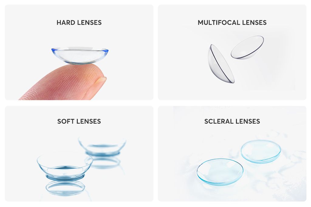
Every patient is different and so are their eyes. This means that there need to be different types of contact lenses to suit each individual. Some patients have corneal abnormalities which mean that conventional lenses won’t sit comfortably on the surface of their eyes, while others suffer from eye conditions that mean normal contact lenses won’t be comfortable or could irritate their eyes.
As you may have guessed from the name, specialty contact lenses are unconventional contacts that are designed for patients that regular contacts might not be suitable. Here are some of the main types of speciality contact lenses and who they are recommended for.
Who might be a good patient for specialty contact lenses?
Some of the patients that might benefit from specialty contact lenses include those who:
have been diagnosed with dry eye syndrome
have corneal scarring
have been diagnosed with keratoconus, a condition characterized by the bulging of the cornea
suffer from strabismus, a condition where the patient has an eye that turns in or out relative to the other
have suffered an injury to the eye
suffer from a peripheral corneal thinning disorder
are intolerant to other types of lenses
Your eye doctor or contact lens provider will be able to tell you if you need specialty contact lenses and if so, which lenses would be best based on your individual requirements.

Corneal refractive therapy, also known as CRT, is a simple, painless treatment for refractive eye errors like myopia and has two core benefits. First, it can be used to help patients see clearly during the day without using glasses or contact lenses, giving them the freedom and flexibility that they need to live life to the fullest. Second, CRT has been shown to help slow the progression of myopia, keeping prescriptions under control and potentially reducing the likelihood of patients developing serious eye health problems associated with high myopia in the future.
Here’s everything that you need to know about corneal refractive therapy and what it means for you.
Understanding refractive eye problems
Refractive eye problems like nearsightedness, farsightedness and astigmatism are extremely common, with nearsightedness – also known as myopia – being the most common of all. Patients with myopia can see nearby objects clearly, but those further away become progressively more blurred. Refractive eye errors occur when the shape of the clear dome covering the front part of the eye, called the cornea, impair the light-bending and focusing process in your eyes. This leads to the light ending up in the wrong place inside the eye, and the message that is sent to our brain from our eyes is muddled, causing blurred vision.
What is corneal refractive therapy?
Corneal refractive therapy was initially developed as a treatment to correct and slow the progression of nearsightedness. However, it has also been found to be effective at controlling other refractive errors, including farsightedness, astigmatism and an age-related refractive condition called presbyopia.
CRT is a non-invasive, painless and straightforward method of correcting patient vision so that they don’t need to wear contacts or glasses, and they don’t need laser vision correction surgery to see clearly. CRT uses special contact lenses that are worn overnight and apply light pressure to the cornea in order to reshape it so that light is refracted correctly, and the image sent from the eyes to the brain is clear. The cornea is able to retain this new shape even after the contact lenses are removed the next morning, meaning that you can continue to see clearly for several hours. The more consistently you wear your CRT lenses overnight, the longer your eyes will learn to retain their new shape and eventually, patients can enjoy up to 48 hours of clear vision without using prescription lenses. However, the effects aren’t permanent so if you stop wearing the lenses, your vision will gradually return back to normal over the course of a few days.
Slowing the progression of myopia with corneal refractive therapy
Another key benefit of CRT is that it can actually help to slow the progression of myopia. Most people who are nearsighted find that their eyesight gets progressively worse as they get older. This deterioration may not be rapid, but it can end in patients requiring high prescriptions. Studies have found that patients who have high myopia are more likely to develop serious eye problems in the future, including glaucoma, macular degeneration, cataracts and a detached retina. Regular use of your corneal refractive therapy lenses could help keep your prescription stable and lower your risk of developing these problems.
Am I a candidate for corneal refractive therapy?
You may be a candidate for corneal refractive therapy if you:
Have a myopia prescription within specific parameters
Have a prescription for hyperopia, presbyopia or astigmatism within specific parameters
Have stable vision, which means that your prescription hasn’t changed during the last two years
Are not a suitable candidate for laser vision correction
Have a job that makes it impractical or unsafe to wear glasses or contact lenses
Enjoy hobbies that make it impractical or unsafe to wear glasses or contact lenses
Have healthy eyes and are generally in good health
For more information, please contact our friendly and knowledgeable team today.

Neurolens are the first and only prescription lenses that include an element of contoured prism in their design. This prism is designed to bring the patient’s eyes into more equal alignment, and this should help to provide relief from the symptoms that are associated with several eye misalignment conditions, including digital eye strain and binocular vision dysfunction.
What is digital eye strain?
Digital eye strain is the name given to describe a group of symptoms that can occur when someone spends long periods of time using digital devices. Since using digital devices requires the eyes to work harder than normal and we don’t always position our devices the perfect distance away, it can lead to issues such as eye pain, dry and irritated eyes, eye fatigue, light sensitivity and blurred vision. Unsurprisingly, the number of people who are experiencing digital eye strain has grown significantly over the last few years and is expected to continue to do so.
What is binocular vision dysfunction?
Binocular vision dysfunction, also known as BVD for short, is another eye condition but is one that is very misunderstood. Binocular vision dysfunction occurs when the eyes aren’t perfectly aligned, causing your brain and eyes to work harder than normal in order to create a clear visual image and remain focused. This places pressure on the trigeminal nerve, which is the nerve that is responsible for the majority of the sensations that we experience in our head and back. BVD can often manifest as other things owing to the huge range of symptoms that are associated with the condition. These can include, but aren’t limited to:
Blurred vision
Headaches/migraines
Double vision
Motion sickness
Vertigo
Dizziness
Anxiety
Many people don’t think to visit an eye doctor when they are experiencing these symptoms, but all can occur simply because the eyes are out of alignment.
What are Neurolens lenses and how do they help?
As well as containing your usual prescription, Neurolens lenses also contain a specific amount of contoured micro-prism. This micro-prism alters the position of images so that they are aligned in the same plane. This then reduces the pressure on the muscles around the eyes as well as bringing the eyes into alignment, easing the symptoms that the patient has been experiencing.
The amount of prism in Neurolens lenses is decided using the Neurolens eye-tracking device. This non-invasively measures the misalignment that the patient is experiencing, and this is used to form the basis for the patient’s Neurolens prescription. After this, it’s fairly normal for the amount of prism to need to be adjusted by infinitesimal amounts to achieve the optimal relief from your symptoms. Most patients who choose Neurolens treatment see a 50% improvement in their vision as soon as they start to have micro-prism incorporated into their prescription lenses. However, with careful adjustments, many patients see as much as an 80% reduction in the effects of digital eye strain and binocular vision dysfunction.
Want more information about Neurolens? Please contact our knowledgeable eye care specialists.

Contact lenses have become a popular choice for individuals who want to correct their vision without the hassle of wearing glasses. Traditional contact lenses have been around for decades, offering a convenient alternative to eyeglasses. However, advancements in technology have given rise to a new type of contact lens – hybrid contacts.
What Are Hybrid Contacts?
Hybrid contacts are a revolutionary type of contact lens that combine the best features of both soft and rigid gas permeable (RGP) lenses. The rigid center corrects vision by providing precise clarity, while the soft skirt offers comfort and stability. This unique combination allows for the benefits of both types of lenses to be experienced simultaneously.
The central RGP lens of a hybrid contact is made from a rigid material that allows oxygen to pass through to the cornea, ensuring ample oxygen supply to the eyes. This ensures the overall health of the eyes, preventing dryness and reducing the risk of complications associated with limited oxygen flow

All Eye
Care Services

Keep
In Touch
Our Services
Contact Info
Hours of Operation
- Monday 8:30am - 6:00pm
- Tuesday 8:30am - 6:00pm
- Wednesday 8:30am - 5:00pm
- Thursday 8:30am - 5:00pm
- Friday 8:30am - 4:00pm
- Saturday Closed
- Sunday Closed
© 2024 Advanced EyeCare . All rights Reserved. Accessibility Statement - Privacy Policy - Sitemap
Powered by: